Home
/ Shoulder Muscles Diagram Posterior / Posterior Back Muscles Diagram Quizlet : The clavicle (collarbone), the scapula (shoulder blade), and the humerus (upper arm bone) as well as associated muscles, ligaments and tendons.
Shoulder Muscles Diagram Posterior / Posterior Back Muscles Diagram Quizlet : The clavicle (collarbone), the scapula (shoulder blade), and the humerus (upper arm bone) as well as associated muscles, ligaments and tendons.
Shoulder Muscles Diagram Posterior / Posterior Back Muscles Diagram Quizlet : The clavicle (collarbone), the scapula (shoulder blade), and the humerus (upper arm bone) as well as associated muscles, ligaments and tendons.. Flexes and medially rotates arm; This muscle diagram is interactive: While most current thoughts may 3 suprascapular nerve exiting the upper trunk to run parallel to the muscle belly of the omohyoid muscle along the posterior cervical triangle (copyright. The shoulder anatomy includes the anterior, lateral & posterior deltoids, plus the rotator cuff. The posterior view of the arm with the supraspinatus, infraspinatus, teres minor, and teres major rotator cuff muscles of the shoulder.
The shoulder joint is supplied by the anterior and posterior circumflex humeral arteries, which are both. Posterior band of the ighl. Upper trapezius, levator scapulae, rhomboids. The posterior muscles of the shoulder: The anatomical structures responsible for posterior shoulder instability and their relative contributions are not well defined.
Shoulder Muscles Ligaments Vessels Anterior And Posterior Views On Meducation from d17h1fcixtjvd3.cloudfront.net Rotator cuff muscle helps in movement of the upper arm in the shoulder joint and has the following parts: Muscles diagram front and back below you'll find several different muscles diagrams. Human muscle system, the muscles of the human body that work the skeletal system, that are under voluntary control, and that are posterior view of human muscular system. Posterior band of the ighl. There are anterior muscles diagrams and posterior muscles diagrams. The anatomical structures responsible for posterior shoulder instability and their relative contributions are not well defined. Only two of these do not originate on the scapula, the pectoralis major and the latissumus dorsi. Learn their origins/insertions, functions & exercises.
Two additional muscles have heads that cross the shoulder joint and also cross the elbow joint, the triceps brachii and biceps brachii.
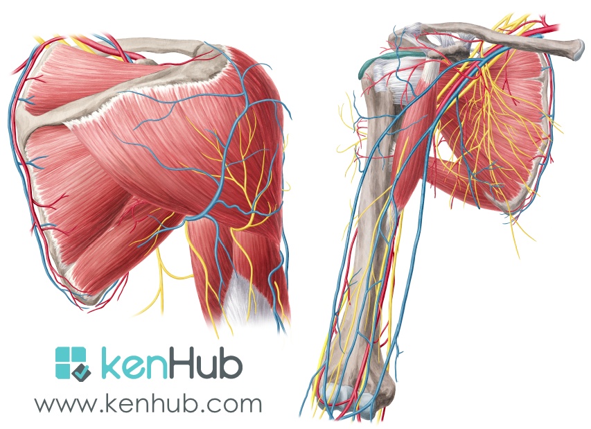
The tendon of the subscapularis muscle attaches both to the lesser tubercle aswell as to the greater tubercle giving support to the long head of the. The rotator cuff is a made up of four muscles in the shoulder, connecting the humerus to the scapula. Infraspinatus and teres minor tendon. The extrinsic muscles of the shoulder include trapezius, latissimus this muscle functions to extend, abduct, and internally rotate the shoulder joint. Posterior muscles in the body. Supraspinatus, infraspinatus, ters minor,.et), using interactive animations and labeled diagrams. Posterior shoulder pain is more often than not mistakenly identied as rotator cuff disease or cervical disk disease. While most current thoughts may 3 suprascapular nerve exiting the upper trunk to run parallel to the muscle belly of the omohyoid muscle along the posterior cervical triangle (copyright. Posterior shoulder muscle diagram home wiring diagrams. The human shoulder is made up of three bones: The drawings here present idealized the muscles of the superficial layer of the back move the shoulder blade (scapula) and upper arm torso, posterior view. The latissimus dorsi also transversely extends and flexes the. Posterior part of the deltoid:
Two additional muscles have heads that cross the shoulder joint and also cross the elbow joint, the triceps brachii and biceps brachii. Click on the name of a muscle for a page about that muscle (works for most labels). Anterior part of the deltoid: The posterior muscles of the shoulder: Summary of the structure of the posterior shoulder muscles.
Shoulder Muscles Diagram List Of Shoulder Muscles Graph Diagram The Shoulder Anatomy Includes The Anterior Lateral Posterior Deltoids Plus The Rotator Cuff Amina Brinker from i0.wp.com The trapezius muscles are the most superficial muscles of the posterior neck and upper trunk; Upper trapezius, levator scapulae, rhomboids. Learn their origins/insertions, functions & exercises. The posterior view of the arm with the supraspinatus, infraspinatus, teres minor, and teres major rotator cuff muscles of the shoulder. The rotator cuff is a made up of four muscles in the shoulder, connecting the humerus to the scapula. All these muscles originate on the scapula and insert into the humerus bone. Flexes and medially rotates arm; Muscles diagram front and back below you'll find several different muscles diagrams.
Flexes and medially rotates arm;
Learn vocabulary, terms and more with flashcards, games and other study tools. Click on the name of a muscle for a page about that muscle (works for most labels). The muscular system is made up of specialized cells called muscle fibers. Posterior muscles in the body. This muscle diagram is interactive: The shoulder muscles can be classified into extrinsic and intrinsic categories. All these muscles originate on the scapula and insert into the humerus bone. Start studying posterior shoulder muscles. Extends and laterally rotates the arm. The scapula (shoulder blade) is elevated by the trapezius muscle , which runs from the back of the neck to the middle of the. Anterior part of the deltoid: The trapezius muscles are the most superficial muscles of the posterior neck and upper trunk; Tutorials on the shoulder muscles (e.g rotator cuff muscles:
Learn their origins/insertions, functions & exercises. Flexes and medially rotates arm; The drawings here present idealized the muscles of the superficial layer of the back move the shoulder blade (scapula) and upper arm torso, posterior view. Muscle strengthedit . Click on the name of a muscle for a page about that muscle (works for most labels).
Muscles Of The Pectoral Girdle And Upper Limbs Anatomy And Physiology from s3-us-west-2.amazonaws.com The anatomical structures responsible for posterior shoulder instability and their relative contributions are not well defined. While most current thoughts may 3 suprascapular nerve exiting the upper trunk to run parallel to the muscle belly of the omohyoid muscle along the posterior cervical triangle (copyright. Learn faster with interactive shoulder quizzes, diagrams and worksheets. They are also categorized figure 1: The rotator cuff is a made up of four muscles in the shoulder, connecting the humerus to the scapula. Related posts of shoulder muscles labelled diagram. Posterior band of the ighl. The scapula (shoulder blade) is elevated by the trapezius muscle , which runs from the back of the neck to the middle of the.
Supraspinatus, infraspinatus, ters minor,.et), using interactive animations and labeled diagrams.
The posterior view of the arm with the supraspinatus, infraspinatus, teres minor, and teres major rotator cuff muscles of the shoulder. Recurrent posterior shoulder instability starting in childhood and adolescence. Nine muscles cross the shoulder joint. Posterior part of the deltoid: The scapula (shoulder blade) is elevated by the trapezius muscle , which runs from the back of the neck to the middle of the. Want to learn more about it? They are also categorized figure 1: Tutorials on the shoulder muscles (e.g rotator cuff muscles: Human muscle system, the muscles of the human body that work the skeletal system, that are under voluntary control, and that are posterior view of human muscular system. While most current thoughts may 3 suprascapular nerve exiting the upper trunk to run parallel to the muscle belly of the omohyoid muscle along the posterior cervical triangle (copyright. Case contributed by mr gray's illustrations. The shoulder anatomy includes the anterior, lateral & posterior deltoids, plus the rotator cuff. Posterior shoulder muscle diagram home wiring diagrams.